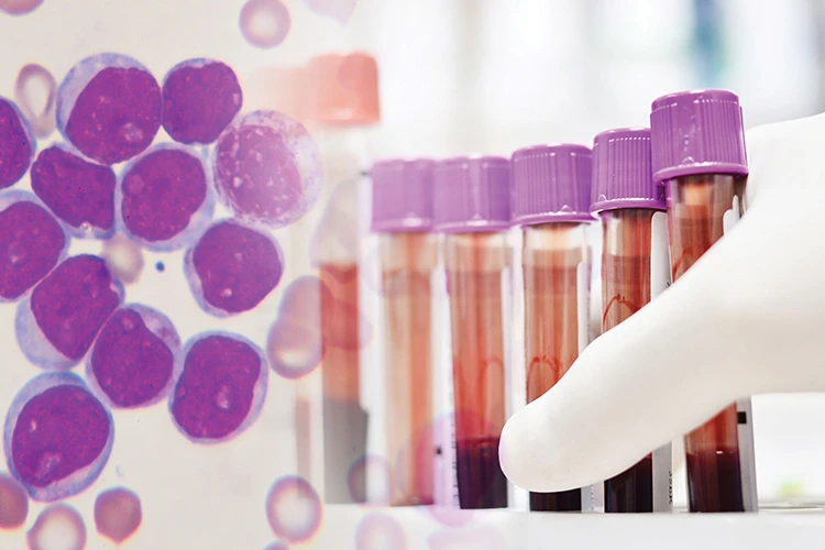
Acute Myeloid Leukaemia (AML)

Acute Myeloid Leukaemia (AML) is a cancer of the blood and bone marrow and is the most common type of acute leukaemia in adults. It causes an overproduction of abnormal myeloblasts (a type of white blood cell), crowding the bone marrow and preventing it from producing normal blood cells. This results in insufficient normal red blood cells, white blood cells and platelets circulating in the body.
AML may also be called Acute Myelocytic Leukaemia, Acute Myelogenous Leukaemia, Acute Granulocytic Leukaemia, or Acute Non-Lymphocytic Leukaemia.
AML may be caused by damage to the genes controlling blood cell development. Certain risk factors for AML have been identified:
- Gender – AML occurs more frequently in men than in women.
- Congenital disorders – People with certain congenital disorders, such as Down Syndrome and Fanconi anaemia, may have a higher risk of developing AML. The AML tends to develop during childhood or adolescence, for such people.
- Pre-existing blood disorders – Certain blood disorders, such as Myelodysplastic Syndromes (MDS), myelofibrosis and aplastic anaemia, may predispose patients to AML.
- Cancer treatment – Certain chemotherapy drugs or radiation therapy may increase the likelihood of developing AML.
- Exposure to chemicals – Long-term exposure to industrial chemicals such as benzene, certain cleaning products, detergents and paint strippers, as well as tobacco smoke, may increase the likelihood of developing AML.
- Exposure to radiation – Exposure to high doses of radiation (for example, from an atomic bomb blast or nuclear reactor accident) increases the risk of developing AML.
AML develops rapidly, so patients may only experience symptoms for a few days to a few weeks before being diagnosed. Some patients do not have symptoms at all. The main symptoms are caused by having insufficient normal blood cells in the body. Common symptoms of AML are:
- Fatigue
- Fever
- Pale skin
- Breathlessness
- Loss of appetite
- Weight loss
- Night sweats or excessive sweating
- Bleeding gums or nosebleeds
- Bruising easily
- Red or purple spots on the skin
- Bone and joint pain
If AML is suspected, a physical examination, blood test and bone marrow biopsy will help in giving a diagnosis.
In the physical examination, the doctor will check for swelling in the liver, spleen and lymph nodes, look for unusual bleeding or bruising, and signs of infection.
The blood test, called a full blood count, involves a sample of blood being sent to the laboratory for investigation. The blood sample will be checked for the number of red blood cells, white blood cells and platelets. A high proportion of white blood cells may indicate AML.
A bone marrow biopsy involves taking a sample of bone marrow, usually from the hip bone. This is done under local anaesthetic and takes 15 – 20 minutes. The sample will also be sent for investigation by the laboratory, to be examined for cancerous cells.
Further tests may be done to determine the extent of the AML and to help determine the best treatment options. Some of these tests are lumbar puncture (or spinal tap), genetic testing, and imaging tests (X-rays, scans and echocardiograms).

For most cancers, staging or determining the stage of cancer is helpful in deciding on treatment options. AML, however, starts in the bone marrow and is generally only detected after it has spread to other organs. Rather than using the traditional cancer staging, AML is classified based on a cellular system.
The French-American-British (FAB) classification classifies AML into eight subtypes based on the number of healthy blood cells, the size and number of leukaemia cells, chromosomal changes in the leukaemia cells and other genetic abnormalities:
- M0 – Undifferentiated Acute Myeloblastic Leukaemia
- M1 – Acute Myeloblastic Leukaemia with minimal maturation
- M2 – Acute Myeloblastic Leukaemia with maturation
- M3 – Acute Promyelocytic Leukaemia
- M4 – Acute Myelomonocytic Leukaemia
- M4 eos – Acute Myelomonocytic Leukaemia with eosinophilia
- M5 – Acute Monocytic Leukaemia
- M6 – Acute Erythroid Leukaemia
- M7 – Acute Megakaryoblastic Leuk
The newer World Health Organization (WHO) classification divides AML into broad groups:
- AML with certain genetic abnormalities
- AML with myelodysplasia-related changes
- AML related to previous chemotherapy and radiation
- AML not otherwise specified
- Myeloid sarcoma
- Myeloid proliferations related to Down Syndrome
- Undifferentiated and biphenotypic acute leukaemias – These are not strictly AML, but are leukaemias that have both lymphocytic and myeloid features. They are sometimes called Mixed Phenotype Acute Leukaemias (MPALs).
The genetic and mutation profile of AML is important for treatment planning and to assess whether patient will benefit from targeted treatment. One of the commonly check mutation is FMS-like tyrosine kinase 3 (FLT-3). FLT-3 inhibitors are promising target, usually used in combination with chemotherapy to improve the outcomes of patients.
Genetic profile can also predict whether a patient needs consolidative treatment with bone marrow transplant or can be treated with chemotherapy alone.
The goal of treatment for AML is to put the cancer into remission and ensure it stays that way. There are two phases of treatment for AML: remission induction therapy and post-remission therapy.
Remission Induction Therapy
Chemotherapy is used to destroy the cancer cells in the body. Anti-cancer drugs may also be given for a subtype of AML called Acute Promyelocytic Leukaemia (APL).
If chemotherapy is successful, the bone marrow should begin making healthy blood cells again.
Post-Remission Therapy
Further chemotherapy may be given to prevent the cancer cells from returning.
A stem cell transplant may be done to help the body regenerate normal bone marrow cells. The stem cells may come from a donor, who are usually family members or voluntary stem cells donors.
Radiation therapy may also be used, in which high-energy X-rays are used to destroy cancer cells in the body. External radiation therapy is provided by a machine outside the body, while internal radiation therapy places a radioactive substance sealed in needles or catheters directly into or near the site of the cancer.
In targeted therapy, drugs or other substances target specific cancer cells to destroy or block their growth, while leaving normal cells unharmed. Clinical trials are still ongoing for various targeted therapies.
When AML is detected early and treated promptly, there is a high probability of remission. A patient is considered to be in remission once there are no longer any signs and symptoms of AML.
Although there is no known way to prevent AML, the following may lower the risk of developing AML:
- Do not smoke.
- Avoid or limit exposure to industrial chemicals such as benzene, such as by wearing protective gear.
- Avoid or limit exposure to radiation, such as by wearing protective gear.
- Avoid treating cancer with radiation and chemotherapy drugs linked to an increased risk of AML. However, some people may require these specific drugs for treatment.

CanHOPE is a non-profit cancer counselling and support service provided by Parkway Cancer Centre, Singapore. CanHOPE consists of an experienced, knowledgeable and caring support team with access to comprehensive information on a wide range of topics in education and guidelines in cancer treatment.
CanHOPE provides:
- Up-to-date cancer information for patients including ways to prevent cancer, symptoms, risks, screening tests, diagnosis, current treatments and research available.
- Referrals to cancer-related services, such as screening and investigational facilities, treatment centres and appropriate specialist consultation.
- Cancer counselling and advice on strategies to manage side effects of treatments, coping with cancer, diet and nutrition.
- Emotional and psychosocial support to people with cancer and those who care for them.
- Support group activities, focusing on knowledge, skills and supportive activities to educate and create awareness for patients and caregivers.
- Resources for rehabilitative and supportive services.
- Palliative care services to improve quality of life of patients with advanced cancer.
The CanHOPE team will journey with patients to provide support and personalised care, as they strive to share a little hope with every person encountered.
